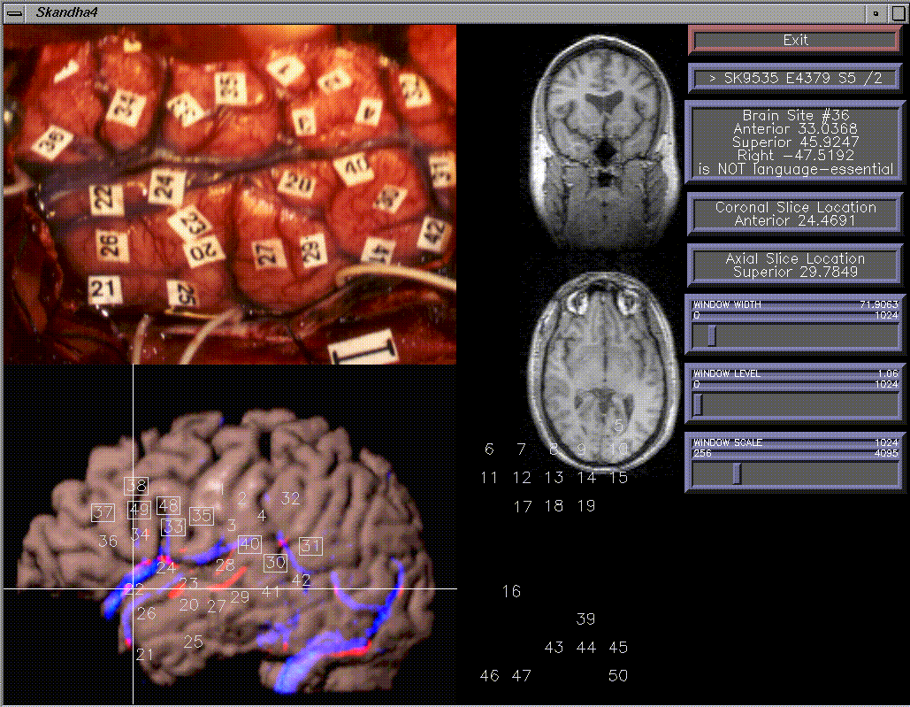
The goal of this research is to develop a framework for organizing and visualizing language mapping data in terms of a 3-D neuroanatomical model. The initial data are cortical electrical stimulation maps, like that shown in the upper left, obtained at the time of neurosurgery for tumors or intractable epilepsy. During the stimulation process some of the sites are designated as essential for language because they interfere with an object naming task. Since the location of these sites varies mapping must be done on each patient in order to plan the resection. The resulting maps become a rich source of information for language models.
In order to correlate the stimulation sites with other data they are first related to a 3-D model of the patient's own brain. The model is reconstructed from MR images acquired prior to surgery, as shown in the bottom left, where the lines indicate the corresponding coronal and transverse MR slices as shown in the middle region. The overall slide is a screen capture of the Brain Mapper tool, which is used to visually match these two images, then to drag numbers from a palette onto the rendering in order to establish the correspondence. The resulting 3-D map coordinates are saved in a database for later retrieval.


