Brain Map Authoring
The first requirement for brain mapping is to relate the surgical sites to a patient-specific 3-D anatomical model. Our current approach is to create a 3-D surface model of the brain from MR scans taken prior to surgery, to render the surface and associated vessels, and to visually match the numbered tags seen on the photograph with the rendered image. The rendering shows cortical arteries and veins, as well as the surface anatomy, since vessels are important landmarks for the surgeon.
Given this approach, the basic problem is to segment the cortex from the MR dataset. Unlike the the anatomy information system, it is not feasible to manually segment the brain for each patient, since we expect hundreds of patients to be included in the study. Therefore, our current segmentation approach in this case is semi-automatic: adaptive 3-D region growing, 3-D mathematical morphology, and volume or surface rendering [Modayur1996, Modayur1997].
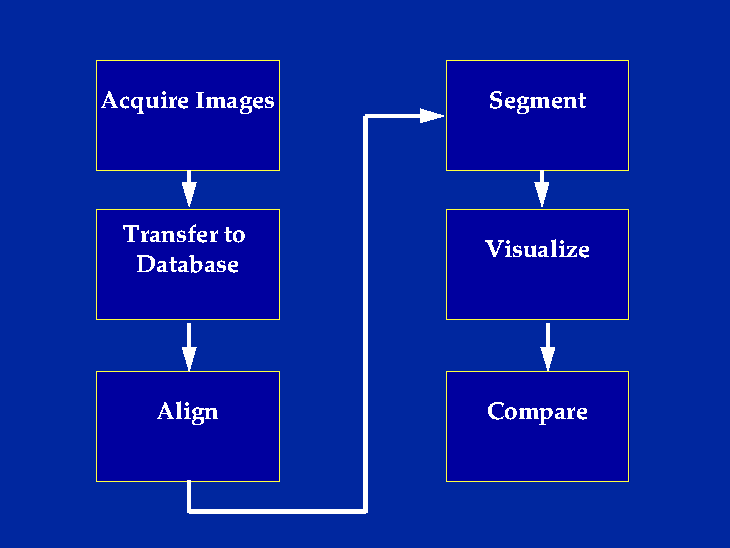
The process is implemented in six stages. Prior to surgery, three sets of MR image volumes are acquired within an interval that is short enough for the patient to remain motionless. One set is optimized to show cortical anatomy, one to show veins, and one to show arteries. The three image sets are transferred over the network from the radiology MR machine to a database in the structural informatics group. They are then aligned by registering and resampling the three datasets within the MR machine coordinate system, so that all voxels correspond and are the same size. The assumption in this case is that the patient does not move during the three sets of image acquisitions.
The cortical dataset is segmented using an interactive 3-D region growing method [Myers1995], implemented in the commercial package AVS. The 3-D region is used as a mask for a standard marching cubes algorithm that extracts the cortex and surface vessels as polygonal meshes. The resulting renderings are the input to the Mapper program for visualization and comparison with the intra-operative photograph.
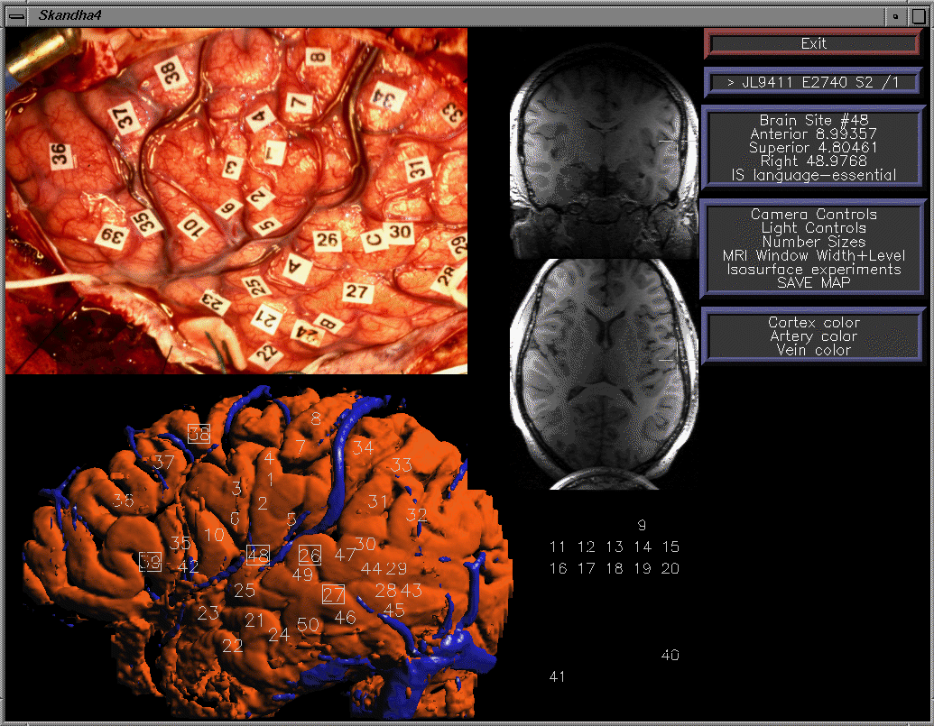
In the Brain Mapper program, the intra-operative photo is shown in the top left, a surface rendering of the segmented cortical, veins and arteries is in the bottom left, and corresponding MR slices are shown in the middle. The surgeon or technician visually matches the two images, then drags numbers from a palette to locations on the rendering that correspond to the photo. Essential sites are indicated by a double click, which causes a box to be drawn around the number. The resulting map is saved in the spatial database as a set of 3-D coordinates with respect to the MR machine coordinate system.