Structural Informatics Group
Digital Anatomist Information System
Integrating spatial and symbolic anatomical knowledge in an online system that permits re-use of anatomical information in multiple applications.
Architectural framework
Example framework diagram. The overall architecture is Internet based, and consists of client applications that access anatomical information resources by means of a set of structural information servers. The exact applications and resources are constantly evolving, but the basic architecture has remained the same for over ten years.2-D annotated images
Java/Swing version of online atlases - Thorax atlas as above, accessed by a Java/Swing client. Includes experimental quiz modes not avaialble in the forms-based atlases: a 2-D puzzle, where pieces of the images are scrambled and the task is to put them back together, and a "Who Wants to Be an Anatomist" game, where the amount of your winnings changes depending whether you point to the correct structure. Jakob Skott and Therese Storheden, visiting CS MS students from Sweden, Fall-Winter, 1999-2000.
Illustration
Non-photo realistic rendering techniques are used to render anatomical scenes so as to resemble medical illustrations. Currently this functionality is implemented by a set of lisp functions that are added to the graphics server that also make use of the 2-D Gimp image manipulation package. A goal is to create an illustrators toolkit that can be added to the scene generator.
Kevin Hinshaw thesis chapter that describes this work.
Coronary Artery Illustration - Some sample images generated from the coronary artery model to explore aspects of automated medical illustration. Also see other examples of general medical illustration. Kevin Hinshaw.
UW Human Brain Project
Using anatomy to organize information about the brain.
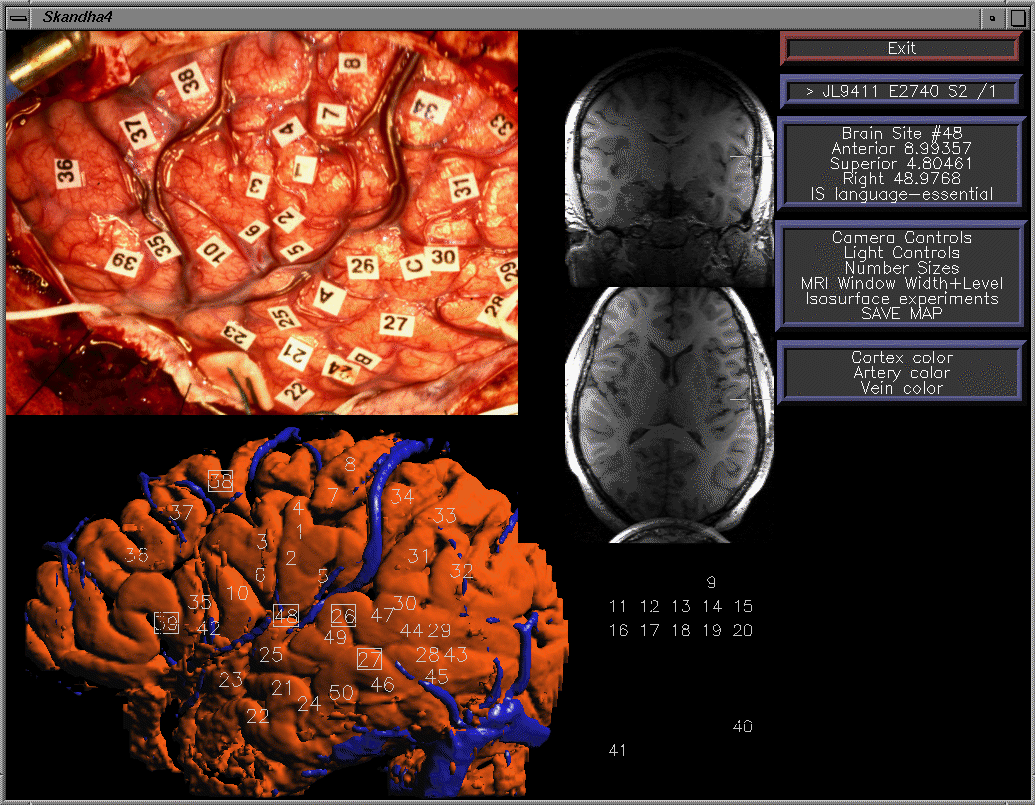
Brain mapper (offline demo) - Interactive mapping of electrical stimulation sites obtained at neurosurgery, onto a 3-D MR-based model of the patient's brain. Kevin Hinshaw and Jeff Prothero.
Brain visualizer (offline demo) - 3-D visualization of multimodality brain mapping data. Andrew Poliakov.